Canon charts course from AI-assisted imaging to AI-assisted diagnosis

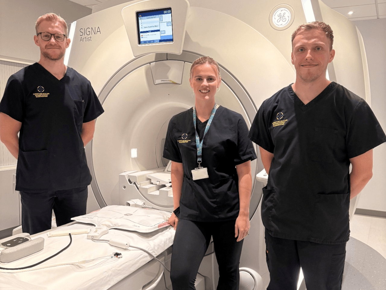
Canon Medical UK has introduced AI-assisted imaging, firstly through CT and now MRI, using the deep learning reconstruction AI algorithm called Advanced intelligent Clear-IQ Engine (AiCE). This differentiates noise from true signal to clean up images resulting in scans free from distortion. It is now used by dozens of UK hospitals.
Improved diagnostic accuracy is cited as one of the benefits of using AI-assisted CT at NHS Lothian’s Royal Hospital for Children and Young People in Edinburgh. Along with making complex paediatric examinations easier, it speeds up scans and reduces dose for young patients. The department’s CT radiographer commented: “Dose reduction for paediatric patients is amazing. The 16cm detector can achieve a volume scan in 0.5 seconds on a head, which is really helpful when examining young patients – we no longer need anaesthetics or strategies to try and keep them still for as long.”
Canon Medical UK has started rolling out AI and automation applications for imaging diagnosis that focus on specific disease conditions. For example, the automation of stroke diagnosis using AI in diagnostic imaging has the potential to streamline stroke-related workflow by automatically consolidating results into a single summary and alerting for abnormalities. AUTOStroke can help to quickly analyse and categorise images to detect signs of ischaemic and haemorrhagic stroke in minutes.
Managing director Mark Hitchman said: “Canon Medical Systems UK is proud to have a home-grown hub of AI research and development via Canon Medical Research Europe based in Edinburgh. This group of software engineers and architects collaborates with 15 partners in the UK across academia, the NHS and industry via its Safe-Haven Artificial Intelligence Platform. This means, in essence, that the data being used to develop future AI innovations is gathered from UK-specific data sources, making the development accurate and specific to our patient populations, at the same time as working closely with colleagues in Japan, China, Europe and the USA.”
Picture: Left group: AI-assisted CT demonstrating excellent visualisation of detail in small vessels enabled by low noise and high resolution characteristics. The patient was hypertensive with atypical chest pain. Helical gated chest CTA was conducted to rule out acute coronary syndrome and/or aortic dissection.
Right group: Carotid angiography using AI-assisted CT. Low dose ultra helical CT angiogram of the carotids and circle of Willis for stroke workup. AI-assisted CT provides clear visualisation of contrast-enhanced vessels and surrounding soft tissue for confident ruling out of occlusion.
Published on page 20 of the August 2022 issue of RAD Magazine.