Second big bore scanner allows new ways of working at Singleton Hospital

Singleton Hospital in Swansea has acquired a second Philips Big Bore RT scanner with improved 4DCT reconstruction times that help improve the imaging department’s clinical workflow. Radiographers will no longer need to wait until the end of the day to reconstruct a 4DCT scan.
Linked to Philips’ Pinnacle3 RT planning system, the system facilitates the acquisition of images with less noise, while lowering doses to patients. It also comes with O-MAR software, which helps to improve image quality and planning accuracy for patients with metal inserts.
According to members of the radiotherapy and radiotherapy physics teams at Swansea, the scanner will work with additional systems to enable them to introduce several new ways of working. The link between Philips’ TumorLOC and the LAP Dorado 5 laser system means the teams will be able to mark on the patient where they would like the treatment radiographers to set up to the plan isocentre. Communication with the C-RAD Sentinel SGRT system will enable radiographers to acquire 4DCT and DIBH scans without additional respiratory tracking devices, and the functionality of TumorLOC will allow radiographers to undertake outlining and beam application tasks as soon as the patient gets off the bed.
Team members said: “Scanning time is decreased and scan doses to patients are lower than with our previous scanner. The installation went smoothly; we found Philips helpful and it responded quickly to any issues we had.”
Philips’ Big Bore RT is designed as a CT simulator for radiation oncology and therapy. It comes with advanced tools to facilitate accurate, efficient patient marking and simulation workflow. With 60cm true scan field-of-view for full, automatic visualisation, it provides spatial positioning accuracy of less than 2mm between the imaging plane and the laser marking plane for confidence in patient marking.
C-RAD Sentinel is a laser-based optical surface scanning system with the functionality for 4D CT reconstruction and gated imaging. It also provides reference images for patient position and intra- fraction motion detection.
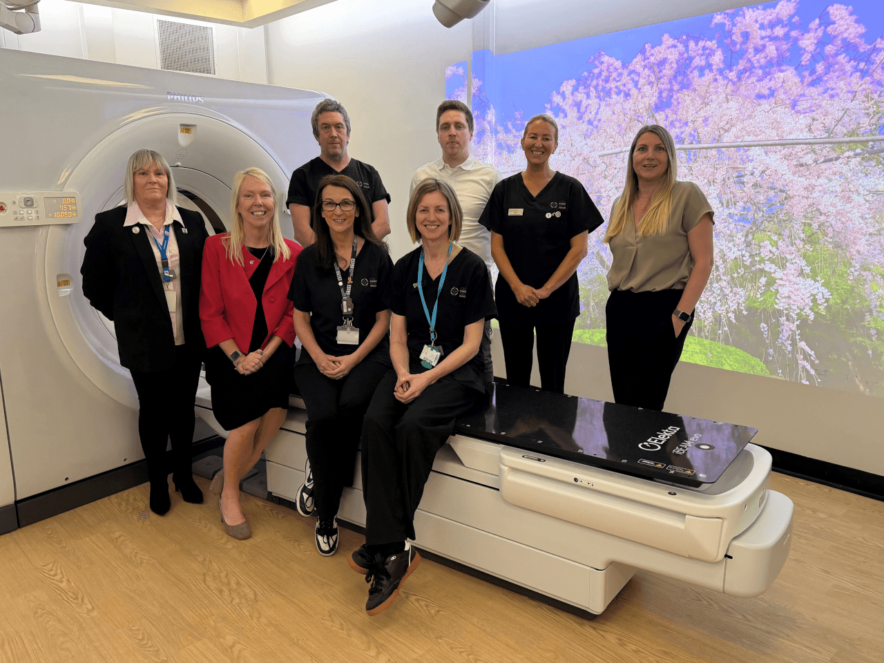
Picture: Philips UK clinical applications specialist – CT Gill Millen, clinical scientist Adam Selby, Prosoma lead radiographer Christine Sillman and CT lead radiographer Helen Streater.
Published on page 20 of the August 2021 issue of RAD Magazine.