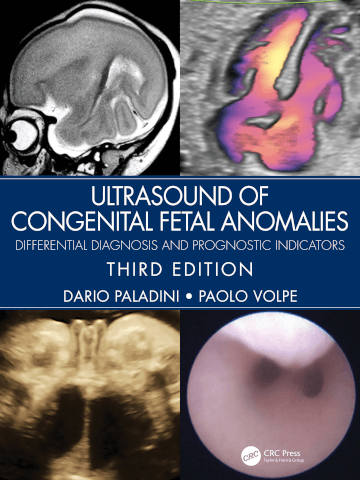
Ultrasound of Congenital Fetal Anomalies: Differential Diagnosis and Prognostic Indicators, third edition
Additional information
|
Author(s):
Paladini, Volpe | Paladini, Volpe |
|
ISBN:
978-0-367-49950-1 | 978-0-367-49950-1 |
|
Publisher:
CRC Press, Taylor & Francis Group | CRC Press, Taylor & Francis Group |
|
Reviewed by:
Ellen Dyer, lead research sonographer, University of Cambridge; Cambridge University Hospitals NHS Foundation Trust | Ellen Dyer, lead research sonographer, University of Cambridge; Cambridge University Hospitals NHS Foundation Trust |
Publisher price: £200
This latest edition of Ultrasound of Congenital Fetal Anomalies is true to its aim of providing clinicians with a comprehensive guide from “ultrasound sign to the final diagnosis”. It features a dedicated section on fetal heart and fetal neurosonography.
The book is up to date and well referenced, reassuring the reader of the latest evidence-based practice in fetal medicine. It is well organised in a logical manner with chapters dedicated to each organ system. This makes it easy to find relevant information, whether during a busy clinical list or for education and learning.
Ultrasound of Congenital Fetal Anomalies would be a useful aid to anyone working in obstetric ultrasound, including sonographers. It does, however, assume some basic understanding of anatomy and ultrasound so is perhaps not suitable for those completely new to scanning. The book is suitable for less experienced ultrasound practitioners who wish to improve their recognition of normal appearances and knowledge of scanning planes.
The book is up to date and discusses many of the most topical issues of modern scanning. There is a strong focus on genetic conditions, and relevant anomalies are cross referenced with the chapter on syndromic conditions discussing numerous syndromes in detail.
Each chapter includes ‘technique boxes’, which are great for practitioners with some experience who want tips on how to optimise their images. These boxes also make it easy to use as a quick reference guide during a busy clinic. In addition, there is also more detailed infor-mation on image optimisation where needed. The fetal heart chapter is a particularly good example of this and provides useful image optimisation suggestions for all levels of practitioner, from trainee to expert.
The images are of high quality and generally well explained. Some of the images are unfortunately missing annotations. This can be overcome by referring to the comprehensive reference list and explanations in the text.
The differential diagnosis flow charts are a useful way to support clinicians as they try to find a cause for a particular ultrasound appearance, for instance the reason for an absent stomach.
For each fetal anomaly, the incidence, ultrasound diagnosis and risk aneuploidy is summarised making the text easy to dip in and out of and directing the reader quickly to the relevant section.
In summary, this book would make an excellent addition to any department’s bookshelf, offering something for everyone.
To purchase this title at our discounted rate email: katherine@radmagazine.com.


