FUJIFILM Sonosite launch 46MHz transducer
FUJIFILM Sonosite is delighted to announce the launch of a revolutionary advancement in point-of-care ultrasound imaging, with the introduction of its UHF46-20 transducer. This groundbreaking transducer is the first and only 46MHz ultra high frequency (UHF) transducer available today in the point-of-care ultrasound market[1], setting a new standard for superficial image clarity and detail.
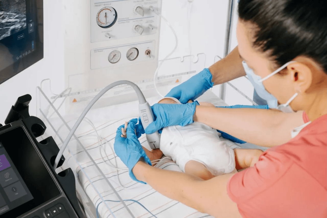
With a minimum scan depth of 4mm, the UHF46-20 transducer will enable clinicians to clearly visualise the first one to two centimetres beneath the skin and identify structures smaller than 1mm, such as superficial nerves and vessels, with high-quality resolution.
Built upon the advanced ultra high frequency technology from FUJIFILM VisualSonics, and available exclusively on the FUJIFILM Sonosite LX POCUS system, this innovative technology provides the best superficial imaging[2,3,4] available on any point-of-care ultrasound system[1].
“At FUJIFILM Sonosite, we are driven by a commitment to help solve real-world challenges clinicians and providers face. The UHF46-20 transducer, when paired with the Sonosite LX system, enables the largest frequency range of any point-of-care ultrasound system on the market today – addressing a long-standing challenge that current clinical ultrasound systems have been unable to overcome,” said Richard Fabian, president and chief executive officer of FUJIFILM Sonosite, Inc. “We’re proud to bring to market the UHF46-20 transducer as the first and only 46MHz UHF transducer in POCUS, to provide clinicians with an unparalleled tool that may help them enhance diagnostic confidence and procedural accuracy.”
The combination of the new transducer and the Sonosite LX offers clinicians a versatile solution for a wide spectrum of imaging needs, spanning from deep abdominal scans to ultra-high resolution superficial assessments. The UHF46-20 transducer holds promise for improving outcomes across a range of sensitive applications including neonatal intensive care unit (NICU) and rheumatology. In the NICU, its superior resolution may help clinicians see superficial submillimetre anatomy that conventional ultrasound may not capture.[6,11] The use of UHF ultrasound may aid clinicians improve procedural quality by allowing for better visualisation of tiny anatomy that may help to improve first attempt success rates.[4,8,9,10]
In rheumatology, UHF’s advanced superficial imaging may aid in the visualisation of subclinical synovitis, erosions, crystal deposits and inflammation, all of which are beneficial in the early diagnosis and intervention of chronic conditions.[2,3,7]
To learn more about the UHF46-20 transducer or to speak to one of our experts click here.
This news story has been sponsored by the companies concerned and does not represent the views or opinions of RAD Magazine.
- Albano, D., Aringhieri, G., Messina, C., De Flaviis, L., & Sconfienza, L. M. (2020). High-Frequency and Ultra-High Frequency Ultrasound: Musculoskeletal Imaging up to 70 MHz. Seminars in musculoskeletal radiology, 24(2), 125–134. https://doi.org/10.1055/s-0039-3401042
- Russo, A.; Reginelli, A.; Lacasella, G.V.; Grassi, E.; Karaboue, M.A.A.; Quarto, T.; Busetto, G.M.; Aliprandi, A.; Grassi, R.; Berritto, D. Clinical Application of Ultra-HighFrequency Ultrasound. J. Pers. Med. 2022, 12, 1733. https://doi.org/10.3390/jpm12101733
- Ait Ichou, J., Gauvin, S., & Faingold, R. (2021). Ultra-high-frequency ultrasound of superficial and musculoskeletal structures in the pediatric population. Pediatric radiology, 51(9), 1748–1757. https://doi.org/10.1007/s00247-021-04978-0
- Hayashi, A., Giacalone, G., Yamamoto, T., Belva, F., Visconti, G., Hayashi, N., Handa, M., Yoshimatsu, H., & Salgarello, M. (2019). Ultra High-frequency Ultrasonographic Imaging with 70 MHz Scanner for Visualization of the Lymphatic Vessels. Plastic and reconstructive surgery. Global open, 7(1), e2086. https://doi.org/10.1097/GOX.0000000000002086
- Latham, G. J., Veneracion, M. L., Joffe, D. C., Bosenberg, A. T., Flack, S. H., & Low, D. K. (2013). High-frequency micro-ultrasound for vascular access in young children–a feasibility study by the High-frequency UltraSound in Kids studY (HUSKY) group. Paediatric anaesthesia, 23(6), 529–535. https://doi.org/10.1111/pan.12131
- Viviano, S. L., Chandler, L. K., & Keith, J. D. (2018). Ultrahigh Frequency Ultrasound Imaging of the Hand: A New Diagnostic Tool for Hand Surgery. Hand (New York, N.Y.), 13(6), 720–725. https://doi.org/10.1177/1558944717731856
- Jacobsen RB, Hebelka H, Gatzinsky V, Elfvin A, Dangardt F. Ultra-high-frequency ultrasound (48–70 MHz) is a promising tool for improved gastrointestinal diagnostics in infants. Acta Paediatr. 2024; 113: 2304–2311. https://doi.org/10.1111/apa.17342
- Brusciano, V., & Lecce, M. (2024). Advantages of the use of ultrasound in newborn vascular access: a systematic review. Journal of ultrasound, 27(2), 203–207. https://doi.org/10.1007/s40477-023-00832-1
- Currie M, Vashisht R, Elkin D, et al. Ultrasound Intravascular Access. [Updated 2024 Jul 2]. In: StatPearls [Internet]. Treasure Island (FL): StatPearls Publishing; 2025 Jan-. Available from: https://www.ncbi.nlm.nih.gov/books/NBK448093/
- Salvia, G.; Zerbinati, N.; Manzo Margiotta, F.; Michelucci, A.; Granieri, G.; Fidanzi, C.; Morganti, R.; Romanelli, M.; Dini, V. Ultra-High-Frequency Ultrasound as an Innovative Imaging Evaluation of Hyaluronic Acid Filler in Nasolabial Folds. Diagnostics 2023, 13, 2761. https://doi.org/10.3390/ diagnostics13172761
- Hawez, T., Evertsson, M., Erlöv, T. et al. The use of ultra-high frequency ultrasound in identifying aganglionosis in Hirschsprung’s disease. Sci Rep 15, 15124 (2025). https://doi.org/10.1038/s41598-025-99897-7