Photon-counting CT system lifts the veil on fine details

Siemens Healthineers reports the successful introduction of the Naeotom Alpha, said to be the world’s first photon-counting CT. Cleared for clinical use in Europe and the USA, the system offers improved resolution and a reduction in radiation dose.
GB&I business manager for CT Russell Lodge explained: “More than 15 years ago, work on photon-counting CT and this clinical vision started at Siemens Healthineers. We always believed in the tremendous clinical value and relentlessly worked on it together with our partners. Today, with the introduction of Naeotom Alpha, we are taking a huge step in furthering patient care in a wide range of clinical domains by effectively showing things impossible to see with conventional CT scans. This required a radical rethinking of practically every technological aspect of CT.”
During its research into photon-counting CT imaging, Siemens Healthineers has filed more than 500 patents related to the technology and collaborated closely with clinical partners to test and validate the clinical capabilities and use cases. Six prototypes have been evaluated and improved on over the years. This year, the company presented the first CT scanner with the new technology, released for clinical use. More than 20 systems have already been installed and are used in clinical routine. So far, over 8,000 patients have been scanned. It has a rotation speed of 250 milliseconds, two x-ray tubes and dual-source detectors.
While conventional CT imaging has reached its technical limitations, says Siemens, photon-counting technology enables great improvements including an increase in resolution and a reduction in radiation dose by up to 45 per cent for ultra high resolution (UHR) scans compared with conventional CT detectors. Photon-counting scans contain more useable data, since this technology directly detects each x-ray photon and its energy level instead of first converting it into visible light as with conventional CT imaging.
These aspects combined open up new capabilities, such as scanning a patient’s lung at a high scan speed and obtaining high resolution images with inherent spectral information without the patient having to hold their breath. This spectral information also helps to identify materials inside the body that can be removed from the image should they obstruct an area of interest. Through the reduction in radiation dose, regular examinations such as lung cancer screenings using CT imaging can become routinely available for larger patient populations.
Head of diagnostic and interventional radiology Professor Thomas Kröncke at University Hospital Augsburg, Germany, has been working with a Naeotom Alpha since April and is very impressed by the initial results: “In oncology, we can break down more precisely which tumour types we are dealing with and thus treat them in a more targeted and effective way. It is like a veil that is now lifting. The new technology is a radical improvement on previous imaging. This will redefine our clinical decision making right from scan one.”
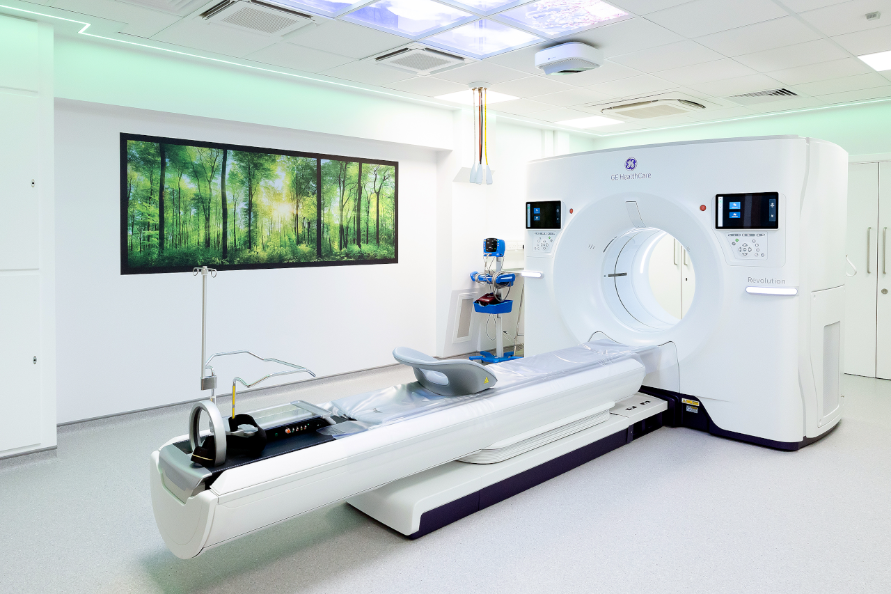
Picture: More than 20 Naeotom Alpha CT systems have already been installed.
Published on page 4 of the December 2021 issue of RAD Magazine.