Success of chest x-ray AI tool leads to introduction of CT Chest by Harrison.ai

Health technology provider Harrison.ai has launched its CT Chest software, an AI tool that assists clinicians in detecting 167 radiological features, including those that may be suggestive of life-threatening conditions, tumours and chronic diseases.
With one of the broadest coverages in chest imaging, CT Chest identifies critical findings such as pulmonary embolism, acute aortic syndrome, pneumothorax and acute rib fractures. According to Harrison.ai, the algorithm prioritises cases on the worklist, intelligently identifies the slices with the findings, localises them with overlays and provides an interpretation with confidence scores, helping clinicians get to time-sensitive cases faster. The tool acts as a safety net, as it is designed to reduce the risk of missed or underdiagnosed conditions, particularly in emergency and inpatient settings where chronic diseases may be overlooked.
The CE-marked software also helps with cancer screening, supporting earlier identification of suspected tumours and lesions across lung tissue, soft tissue, mediastinum, lymph nodes and the upper abdomen. Harrison’s Chest CT assists radiologists by detecting, staging and monitoring features suggestive of lung, gastric and pancreatic cancers, and helping streamline lung screening workflows through time-saving reporting features.
“After the successful market penetration of our chest x-ray AI solution, expanding into CT Chest was a natural next step,” said Harrison.ai co-founder and CEO Dr Aengus Tran. “We’ve seen firsthand how a comprehensive AI solution for a single modality can transform radiology workflows. With CT Chest, we’ve taken the same comprehensive approach to empower clinicians with deeper insights and faster, more confident decision making.”
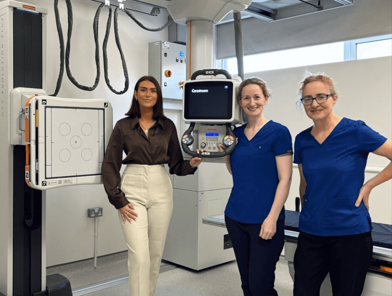
Picture: CT Chest aids the detection of 167 radiological features.
Read this report on page 13 of the December 2025 issue of RAD Magazine.