Transforming breast imaging pathways: the growing role of contrast-enhanced mammography and contrast-enhanced biopsy
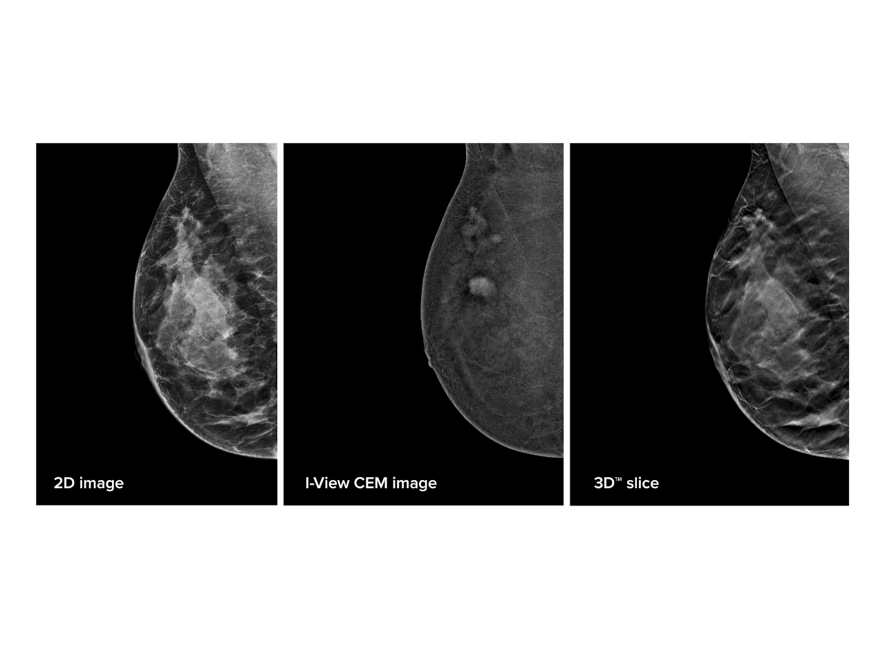
Breast imaging services across the UK are facing unprecedented demand. Rising referral volumes, increasing diagnostic complexity and ongoing workforce pressures are driving the need for innovative approaches that can deliver accurate diagnoses while supporting more efficient patient pathways. As breast services seek solutions that improve both clinical confidence and patient experience, contrast-enhanced mammography (CEM) and contrast-enhanced biopsy are emerging as important technologies with the potential to reshape the diagnostic landscape.
Traditionally, breast cancer assessment has relied on a combination of mammography, ultrasound and, in selected cases, magnetic resonance imaging (MRI). While MRI remains a highly sensitive modality, access can be limited by scanner availability, examination time and patient suitability. CEM offers an alternative approach by combining standard mammographic imaging with the administration of intravenous contrast, enabling clinicians to visualise areas of increased tumour vascularity that may not be apparent on conventional imaging alone.
The growing body of evidence supporting CEM has generated significant interest among breast imaging teams across the UK. Clinicians are increasingly exploring how the technology can support diagnostic assessment, pre-operative planning and the investigation of patients with complex imaging findings. By providing both anatomical and functional information within a familiar mammography setting, CEM has the potential to enhance diagnostic confidence and support more informed clinical decision-making.
One of the most significant developments accompanying the adoption of CEM is the evolution of contrast-enhanced biopsy techniques. In some patients, lesions identified only through contrast enhancement can be difficult or impossible to target using conventional ultrasound or stereotactic guidance. Contrast-enhanced biopsy addresses this challenge by enabling radiologists to directly sample these lesions, creating a pathway to tissue diagnosis without the need for MRI-guided procedures.
This capability has important implications for both patients and healthcare providers. By allowing suspicious findings detected on CEM to be biopsied in the same imaging environment, services may be able to reduce diagnostic delays, minimise the number of appointments required and shorten the interval between detection and diagnosis. For patients, this can mean a more streamlined experience and reduced anxiety during what is often a highly stressful time.
The potential benefits extend beyond individual procedures. As NHS breast services continue to focus on improving efficiency and delivering timely cancer diagnoses, technologies that support faster decision-making and more integrated pathways are attracting considerable attention. The combination of CEM and contrast-enhanced biopsy offers an opportunity to move more seamlessly from detection to diagnosis, helping to optimise utilisation of existing resources while maintaining high standards of care.
Importantly, successful implementation requires careful consideration of local workflows, training requirements and patient selection. As experience with these technologies grows across UK centres, shared learning from early adopters will play a key role in helping organisations understand how best to incorporate CEM and contrast-enhanced biopsy into routine clinical practice and where they can deliver the greatest value.
As breast imaging services continue to evolve, healthcare teams are looking for practical strategies to enhance diagnostic pathways, improve patient experience and support increasingly stretched services. To help healthcare professionals explore these opportunities in greater depth, Hologic recently hosted the webinar, Transforming Breast Imaging Services: The Potential of CEM and Contrast-Enhanced Biopsy on Clinical Pathways.
Led by Dr Tamara Suaris, lead for breast imaging innovation and research at Barts Health NHS Trust, the session shared practical insights into how contrast-enhanced mammography (CEM) and CEM-guided biopsy are being integrated into clinical practice. Drawing on her experience, Dr Suaris explained how “CEM and CEM biopsy is helping us improve our patient pathways with a better and faster experience for patients.”
The webinar is now available to watch on demand.
By registering, healthcare professionals can gain a deeper understanding of the clinical applications of contrast-enhanced technologies, hear real-world experiences from an NHS early adopter, and explore the potential of CEM and contrast-enhanced biopsy to support the future development of breast imaging pathways across the UK.
This news story has been sponsored by the companies concerned and does not represent the views or opinions of RAD Magazine.