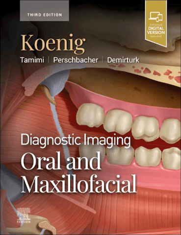
Diagnostic Imaging: Oral and Maxillofacial, third edition
Description
Bridging the gap between dentistry and medical radiology, the third edition of Diagnostic Imaging: Oral and Maxillofacial, is an invaluable resource for anyone who requires an easily accessible, highly visual reference in this complex area of imaging, from new and seasoned radiologists to dental specialists and general practitioners currently using CT and/or cone beam CT (CBCT).
Additional information
|
Author(s):
Koenig, Tamimi, Perschbacher and Demirturk | Koenig, Tamimi, Perschbacher and Demirturk |
|
ISBN:
978-0-443-10531-9 | 978-0-443-10531-9 |
|
Publisher:
Elsevier | Elsevier |
|
Reviewed by:
Dr Calvin Soh, consultant neuroradiologist, Manchester Royal Infirmary | Dr Calvin Soh, consultant neuroradiologist, Manchester Royal Infirmary |
Publisher price: £287.99
Imaging of the oral cavity and maxillofacial region is an entire subspeciality practised by radiologists who cross over between neuroradiology, dental and head and neck radiology. There are very few radiologists who subspecialise only in oral and maxillofacial imaging. Dentists, oral and maxillofacial surgeons may be self-sufficient in interpreting imaging in dental radiology.
This work by Professor Koenig and co-authors is an encyclopaedia that encompasses everything from the suprahyoid neck to the skull base, including the cranial nerves. The book is divided into four parts, covering anatomy, imaging applications in the oral maxillofacial region, diagnoses and differential diagnoses. Each part is then organised into sections based on anatomical site. The section on diagnoses encompasses almost every pathology you can think of, while the part on differential diagnoses helps the reader immensely in clinical practice by organising the thought processes.
The format of this book utilises the same successfully tried and tested formula in other textbooks in the Elsevier series, in the same vein as the popular Diagnostic Imaging: Brain by Anne Osborn, that most neuroradiologists are familiar with. Each condition in the diagnoses section is organised into imaging appearances, the relevant differential diagnoses, the pathology and the clinical issues. In some conditions, there is also a very helpful note on ‘imaging interpretation pearls’ and another on ‘reporting tips’. The information is succinct but detailed. If the amount of information does not impress the reader, the imaging and illustrations will definitely astound them.
The content is huge. For the experienced who are practising this speciality, you can rapidly thumb through the book and consolidate all your knowledge. For neuroradiologists who need a quick reference, the information is easily accessible at your fingertips to reach a diagnosis. The illustrations are so good, one can easily digest this by going through the images and reading around the subject. The anatomical illustrations in the first section on anatomy are superb, with anatomical diagrams, imaging correlates and even 3D reconstructions.
This book is beautifully presented. The downside is its weight. Fortunately, a digital version is available as an accompaniment, which literally provides information at your fingertips.
This is highly recommended for all radiologists. It is a valuable reference text for the experienced practising head and neck radiologist, but comprehensive enough to upskill any novice interested in the subject to improve their clinical practice.
The authors must be congratulated on this excellent masterpiece. It is a work of art.
To purchase this title at our discounted rate email: katherine@radmagazine.com.


