Chest CT AI facilitates identification of nodules to indicate progressive disease

The department of medical imaging at Isala Hospital in Zwolle, the Netherlands, has been using diagnostic AI applications for four years and contextflow’s Advance Chest CT software since the end of 2022. Radiologist Dr Martijn Boomsma has reported on the department’s experience.
Before going live, various issues had to be overcome and implementation was not a plug-and-play process. These issues were not only the vendors, but also internal processes. However, as Dr Boomsma pointed out: “This is where true partnership shows itself and we are still experiencing this with contextflow.” He also noted contextflow’s efforts to continuously develop its solution in close cooperation with its users in order to optimise workflow and increase diagnostic value. “We see a high level of professionalism and agility, as well as the company’s vision to get the best out of AI in thoracic imaging.
“Advance Chest CT helps me identify and quantify nodules. This allows me to clearly identify whether the disease is progressive or stable.” As well as pulmonary nodules, Dr Boomsma uses the AI for the detection and quantification of emphysema. He also sees the potential to speed up the reporting of findings, citing scans without significant findings as an example. The algorithm can mark these as such, so that the radiologist only has to check them once and can then concentrate on more complex cases.
“Advance Chest CT gives me much more information than AI solutions from other vendors. It detects and quantifies nodules, emphysema and also fibrosis. The precise information on the extent of manifestation of suspected pathology adds real value to the findings because it can be indicative and correlate with the patient’s condition, and may guide the need for therapy,” Dr Boomsma continued.
With the search feature an overview of similar cases opens from an extensive database, which radiologists can use to support their diagnosis. The timeline module visualises the changes in detected nodules over time, showing the percentage of growth as well as the time of volume doubling.
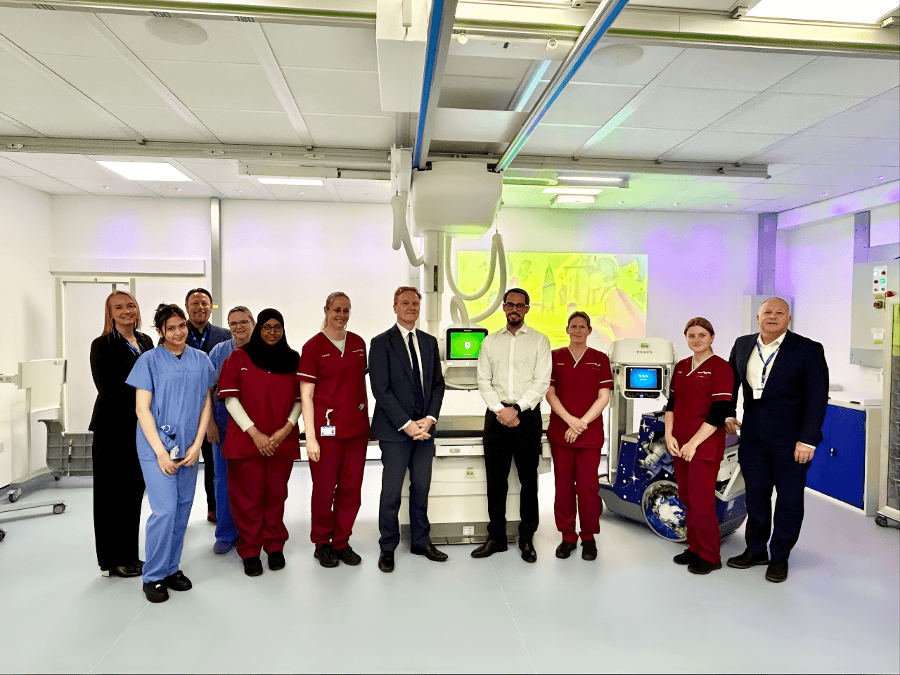
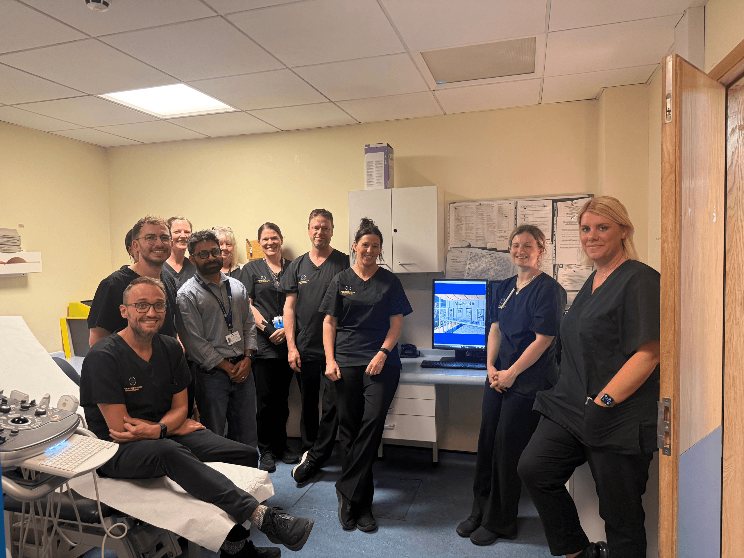
Picture: Isala Hospital’s medical imaging department teamed up with contextflow to implement Advance Chest CT software.
Published on page 21 of the July 2023 issue of RAD Magazine.