Neuroradiology: head, neck and brain trauma – errors and pitfalls in artificial intelligence, machine learning and Chat GTP
The brain, head, neck, and spine are at the forefront of emergencies that occur in medicine, making time of the essence and scans critical for radiologists. Neuroradiology includes a wide range of emergency trauma situations such as strokes, cerebral aneurysms, spine fractures, skull fractures, brain tumors, concussions, contusions and ruptures. Artificial intelligence has the power to completely transform how MRI and CT images are read by neuroradiologists, diagnostic radiologists, and interventional radiologists; in turn transforming actions taken by emergency room doctors, orthopaedic surgeons and trauma surgeons during times of crisis in emergency departments across the world.
By chance, the anatomy of the brain has a highly complex, sophisticated, interdimensional layout, also rendering it necessary for several different modalities of neuroimaging technology to scan for potential issues. This provides a perfect combination and setup for machine learning and deep learning. A type of machine learning specification called radiomics is a medical imaging method that quantitatively employs artificial intelligence to interpret large amounts of highly intensive neurological data. This data may be invisible and regularly missed from standard hospital MRI, CT, and PET brain scans. Radiomics can reveal complex scan texture that the human eye cannot see.
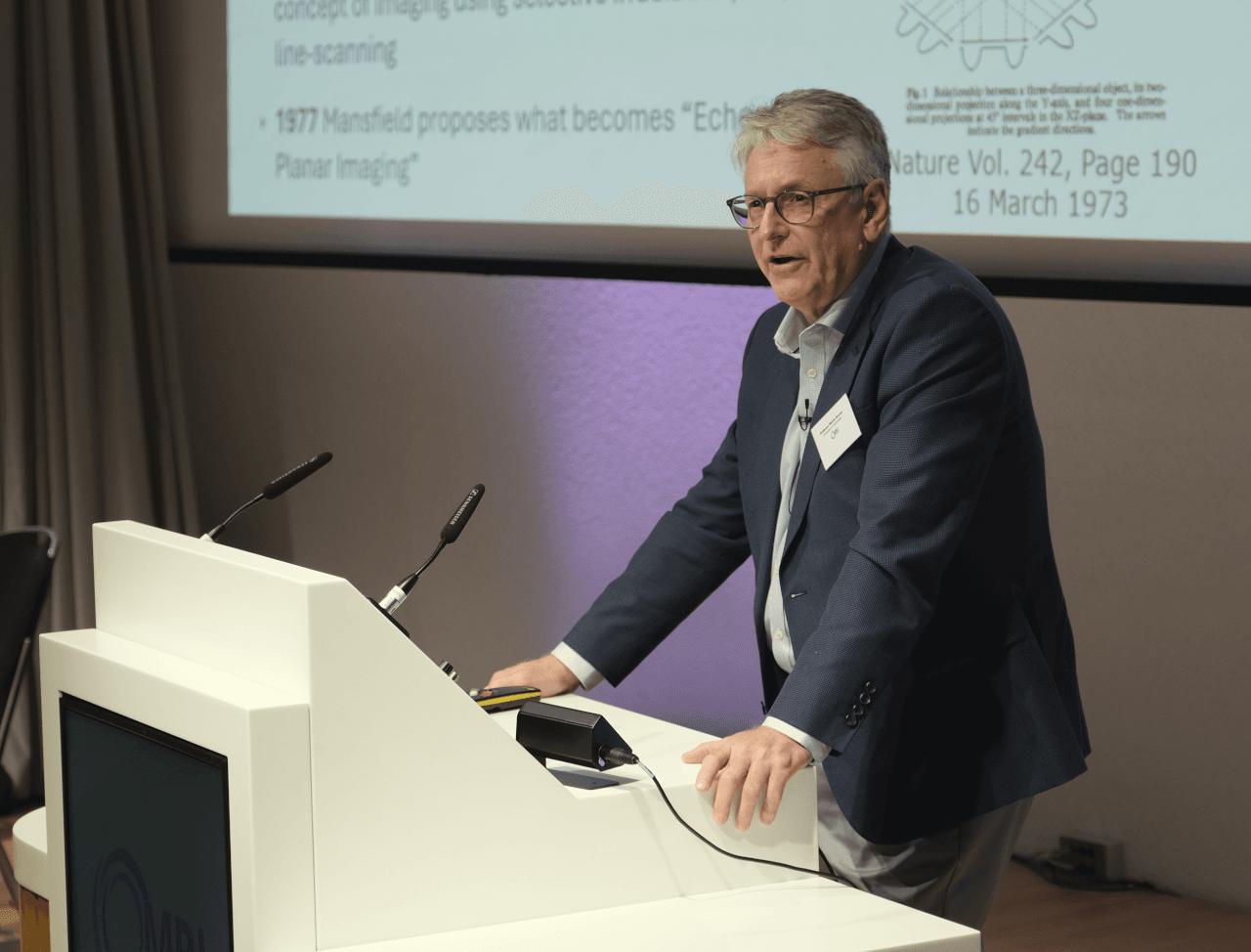
The author of this article, Dr Ana Bhattacharyya, interviewed Dr Daniel Ginat, neuroradiologist and director of head and neck imaging at University of Chicago Pritzker School of Medicine, and CEO of the Chicago neuroimaging company RadLabAI, to get his thoughts on this topic.
How has AI, radiomics and Chat GPT helped and assisted in diagnosing brain conditions, lesions, and trauma?
There are many commercially available products in use that many of us find to be helpful, such those for identifying acute intracranial haemorrhage.
How has AI, radiomics and Chat GPT misdiagnosed, hindered, or hurt in diagnosing brain conditions/lesions/trauma etc?
I do not think these have had any significant detrimental impacts. While these tools are not perfect, they generally have some false positives, which is not necessarily problematic since it is often considered better to identify a potential abnormality than to miss it. Ultimately, it is up to the radiologist to review the scans and make a final decision.
What are the most common dangerous mistakes in diagnosing/misdiagnosing head and neck disorders that you see, potentially leading to injury, coma,or death?
There are many challenges in head and neck imaging. One theme I encountered in medicolegal cases is missed parotid tumors either on head or neck CT’s, especially reviewing the parotid glands on head/brain imaging. In my upcoming book Neuroradiology Law and Order I will include other topics.
Can AI, radiomics or Chat GPT make this misdiagnosing situation even worse?
I suppose that there could be a danger if the software is not trained properly. For example, I have heard that some of the automatically generated impressions sometimes do not emphasise critical findings. Once, again, it is still the responsibility of the radiologist to not only rely on the software, but use their own.
How do you see the future of neuroradiology evolving?
I envision a greater shift towards automation and quantified imaging, which will contribute to personalised medicine and via my company RadLabAI I am excited to be involved in future research and development.
Further reading
1. Daniel Ginat. Implementation Of Machine Learning Software On The Radiology Worklist Decreases Scan View Delay For The Detection Of Intracranial Hemorrhage On CT. Brain Sciences. 2021; 11(7):832. https://DOI.org/10.3390/brainsci11070832
2. Daniel Ginat, Anasuya Bhattacharyya, Mathew Illimmottil. Preoperative And Postoperative CT Imaging Assessment Of Obstructive Sleep Apnea. Journal of Computer Assisted Tomography. 2025 Nov-Dec 01;49(6):985-992. DOI: 10.1097/RCT.0000000000001748. Epub 2025 Mar 20. PMID: 40165025.
3. Daniel Ginat, Ayden Olsen. Assessment Of Commercially Available Artificial Intelligence Software For Differentiating Hemorrhage From Contrast On Head CT Following Thrombolysis For Ischemic Stroke. Front Neurol. 2025 Mar 4;16:1458142. DOI: 10.3389/fneur.2025.1458142. PMID: 40103936; PMCID: PMC11915465.
Submitted by independent consultant Dr Anasuya Bhattacharyya.
The content on this page is provided by the individuals concerned and does not represent the views or opinions of RAD Magazine.