Integration of 3D ultrasound with software assists surveillance of abdominal aortic aneurysms

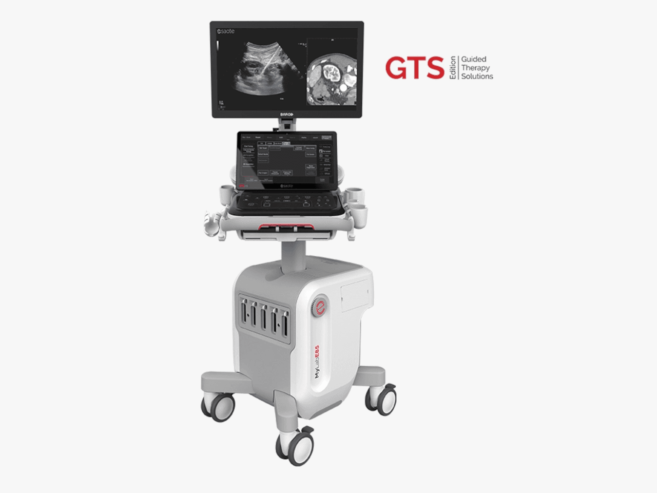
Royal Philips has introduced the Philips Abdominal Aortic Aneurysm (AAA) Model, said to provide a more patient-friendly solution compared to the current standard of care for managing AAA patients. Based on 3D ultrasound, Philips AAA Model delivers accurate diagnostic information without exposing patients to high doses of radiation and nephrotoxic contrast agents.
Typically, AAAs are identified incidentally during abdominal imaging examinations but, in some cases, remain undetected until rupture. The model integrates innovative software and Philips 3D ultrasound technologies to help increase diagnostic confidence and an improved patient experience. The software automatically segments and quantifies the size of the aneurysm sac for surveillance of known native (untreated) and post-EVAR (treated) AAAs.
“A recent clinical study showed that 3D ultrasound examination for native AAA surveillance has excellent inter-operator reproducibility, superior to that of 2D ultrasound, supporting the broader use of 3D ultrasound in standard AAA surveillance programmes,” said Philips. “3D ultrasound has been shown to estimate the diameter and volume of an AAA with acceptable reproducibility and an improved agreement (over 2D ultrasound) with CT. Furthermore, 3D ultrasound has also been proven to correlate significantly better to 3D CT than 2D ultrasound for assessing the maximum diameter of the residual sac post-EVAR, with clinically acceptable reproducibility.
Picture: The Philips AAA Model is CE marked for sale in Europe.
Published on page 12 of the March 2021 issue of RAD Magazine.