Exploring factors influencing TVUS acceptance among ethnic minority and white British women in the UK
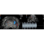
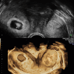
Transvaginal ultrasound (TVUS) is a pivotal diagnostic tool in gynaecology, offering high accuracy in detecting pelvic pathologies. Despite its clinical efficacy, TVUS faces significant acceptance barriers, particularly among ethnic minority women in the UK. This mixed-methods study investigates cultural, informational and gender-based factors influencing TVUS acceptance across diverse ethnic groups. Twelve women from varied ethnic […]

Nargis Begum
Royal Stoke University Hospital